Thanks for spending part of your week with Omic.ly!
There will be some changes around here, namely, you're going to start getting the weekly reading list for free and there will be bonus hot takes from time to time.
Thanks for your continued support!
This Week's Headlines
1) A gene for short sleep duration
2) Structure equals function might be the most important phrase in proteomics
3) Non-invasive prenatal testing is a big business. Here's how it got its start.
4) Weekly Reading List
Having trouble sleeping? A new mutation has been discovered that allows people to feel well rested after as little as 4hrs of sleep!
If you're anything like me, you cherish your sleep!
And also, if you're anything like me, you feel like you can never get enough!
Between the tossing and turning and constant worrying about how everything can go wrong, I'm lucky if I get 6 hours of sleep every night.
But did you know that there are a number of genetic mutations that have been identified that allow people to feel well rested after only getting 4 hours of sleep?!
That's half the recommended dose!
And about 2 hours less than I normally get.
Some people (and mice) have all the luck!
They also likely have mutations in sleep related genes like DEC2, ADRB1, NPSR1, and GRM1.
Mutations in these genes have been shown to be associated with natural short sleep (nss) which allow for short sleep duration without all of the negative cognitive or physiological side effects associated with chronic sleep deprivation.
People with mutations in these genes do not crave more sleep and may even feel worse when oversleeping!
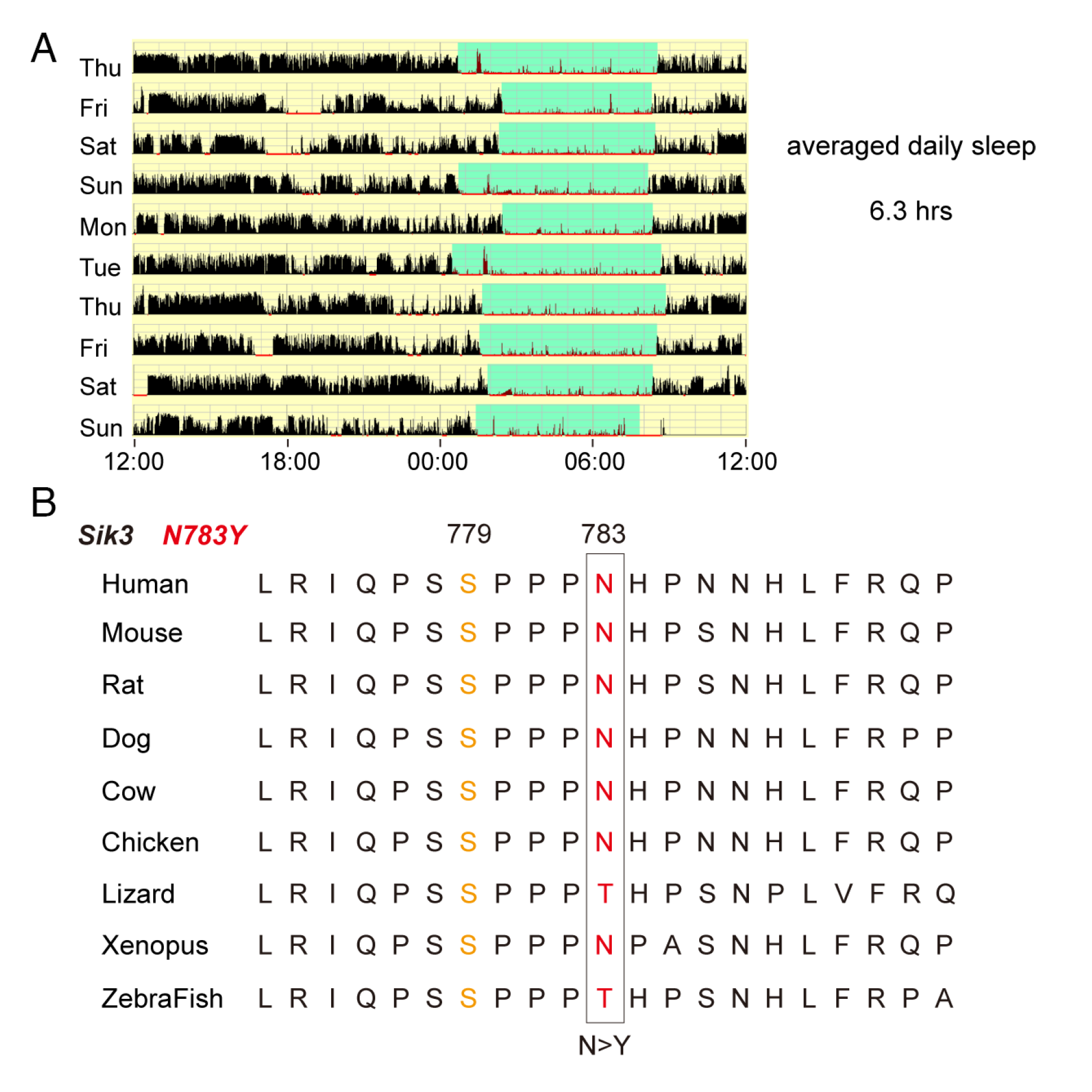
Now, researchers have identified yet another mutation in a gene called SIK3 (Salt-Inducible Kinase 3) which allows those who carry an N783Y mutation all of the benefits of a full night of sleep after only 4-ish hours.
This can be seen in the figure above which displays A) an actogram (activity diagram) of a person who carries this mutation highlighting their sleep duration in green and B) a conservation map of the SIK3 gene across vertebrates (animals with spines).
In mouse models harboring the SIK3 N783Y mutation, the researchers showed that homozygous animals exhibited significantly reduced sleep both under baseline conditions (31 minutes less) and during recovery after sleep deprivation (54 minutes less).
However, the phenotype was milder than in humans, which could just be related to some species specific differences in how SIK3 operates.
Functional assays further revealed that this mutation reduces kinase activity (addition of phosphate groups to other proteins) and that approximately 20% of synaptic phosphoproteins exhibited altered phosphorylation due to the N783Y mutation.
This implicates SIK3 in maintaining synaptic homeostasis (stabilizes neural circuits in your brain), a critical function during sleep.
This study also strengthens the case for SIK3 as an evolutionarily conserved sleep-promoting gene and supports the therapeutic potential of SIK3 as a target for treating sleep disorders.
###
Chen H., et al. 2025. The SIK3-N783Y mutation is associated with the human natural short sleep trait. PNAS. DOI: 10.1073/pnas.2500356122
Proteomics: It’s all about the structures!
‘Structure Equals Function.’
It’s basically the secret password at the molecular biology club.
And if you’re ever looking to get knowing nods from your favorite molecularly aligned friends, this phrase kills.
That’s because most everything in molecular biology is centered on how molecules interact with one another.
This includes how two strands of DNA coil into a right-handed helix, how proteins bind to one another to create functional complexes, or how enzymes grab on to their co-factors and substrates to catalyze life-sustaining chemistry.
All of these things are essentially determined by their molecular structures!
These structures are pretty easy to determine for run of the mill chemical compounds, but proteins are macromolecules.
This just means they’re composed of thousands of atoms and can have highly complex structures!
But the complexity for proteins doesn’t end there, because they can also be modified after they are made.
These ‘post-translational modifications,’ or PTMs for short, can be as simple as the addition of a phosphate to a specific part of a protein all of the way to the addition of large chains of sugar molecules.
And you guessed it, the addition of these PTMs changes the structure of the protein which in turn changes its function!
Many PTMs are critical for proteins to operate correctly and they routinely serve as ‘on’ and ‘off’ switches.
They’re also important for determining which partners a protein can interact with and when!
So, a protein’s structure is REALLY freakin’ important for understanding what it does.
'But how do we determine the structure of a protein?'
I'm glad you asked!
X-ray Crystallography - The OG of structure determination involves creating pure extracts of the protein you want to look at and finding the perfect conditions (salt concentration, humidity, drying time) to create a pristine crystal. Those crystals are then bombarded with X-rays. And the structure of the underlying protein can be inferred based on how those X-rays bounced off of it! But one major drawback of crystallography is that crystals don’t have water! So they may not necessarily represent the ACTUAL structure we’d see inside of a wet environment.
Nuclear Magnetic Resonance Spectroscopy (NMR) - Best used on small proteins and peptides. Purified protein is exposed to radio waves and its structure can be determined by looking at resonance signals and calculating which atoms within a protein are near one another.
Cryogenic Electron Microscopy (CryoEM) - Purified proteins or large protein complexes are cryogenically frozen and then imaged with an electron microscope. The images of thousands of molecules are then combined to stitch together a 3D structure of the protein!
AlphaFold - Kidding, this is a structure prediction tool. It’s important to validate what this produces with one of the above methods.
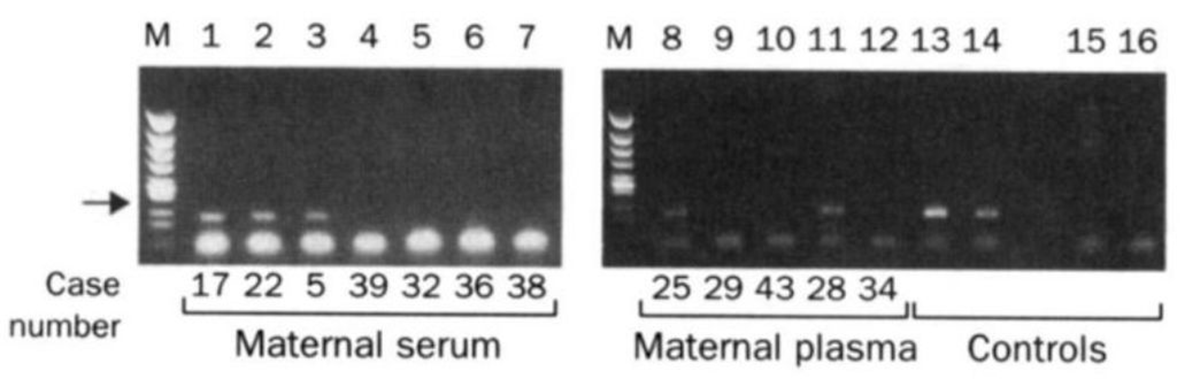
The two gels below spawned a multi-billion dollar industry that didn't exist prior to their publication in 1997.
While it may seem obvious today, you might find it surprising that we didn't know that fetal DNA was present in a pregnant mother's bloodstream until the late 1990's.
Prior to this discovery, genetic testing on fetuses was only performed if a problem was suspected with a pregnancy or if there was a family history of genetic disorders.
This testing was performed by karyotyping fetal cells.
You've probably seen one of these 'chromosome spreads' in a biology textbook where each of the 23 pairs of chromosomes are lined up next to one another.
This allows a geneticist to check that the chromosomes are intact and see if there are any abnormalities such as those found in Down Syndrome where patients have 3 copies of chromosome 21 instead of 2.
But, back in 1997, the process for collecting fetal cells was very invasive and was done using one of two techniques:
Amniocentesis (Amnio) - a 3-5" long needle is inserted into the mother's abdomen to collect 20 milliliters of amniotic fluid for testing.
Chorionic Villus Sampling (CVS) - A 6" long needle is guided via ultrasound, either vaginally or through the abdomen, to obtain tissue from the placenta for testing.
Unfortunately, these procedures carry a risk and 1-2% of the time they can lead to the loss of the fetus.
Recognizing that there had to be a better way, Yuk-Ming Dennis Lo set about figuring out how to get at fetal DNA without having to perform Amnio or CVS.
He knew that fetal cells made it into the maternal bloodstream and that mother and child exchanged cellular material.
But he couldn't isolate enough fetal cells from the blood to do prenatal genetics with them.
Luckily, in 1996, Lo heard that a team in Switzerland had shown that tumors actually shed cell free DNA into the bloodstream and this tumor DNA could be detected using PCR and primers specific to the tumor DNA.
He reasoned that a baby, or more accurately, the placenta, was basically like a giant tumor and shared the bloodstream with the mother.
So, it made sense that it too should shed cell free DNA into the bloodstream.
The figure above is proof that Lo's hypothesis was correct and fetal DNA could be isolated and amplified from the blood of pregnant females.
In this figure, the arrow highlights a 198bp PCR product from the Y chromosome. Case numbers greater than 30 are pregnancies with female fetuses (don't have a Y), and case numbers less than 30 are male fetuses (have a Y).
Further technical advancements and the invention of high throughput sequencers have transformed this discovery into a popular pregnancy screening test.
Today, non-invasive prenatal testing is second only to oncology testing in sequencing market share.
###
Lo YMD, et al. 1997. Presence of fetal DNA in maternal plasma and serum. The Lancet. DOI:10.1016/s0140-6736(97)02174-0