Thanks for spending part of your week with Omic.ly!
This Week's Headlines
1) Nerve cells transfer their mitochondria to cancer cells
2) The microbiome has more to do with traditional ecology than you might think
3) The most beautiful experiment in biology
4) Weekly Reading List
Just when you thought cancer couldn't get any crazier...neurons can directly transfer their power producing mitochondria to tumor cells
Cancer cells, especially metastatic ones, have exceptionally high energy demands.
To survive in changing and often hostile environments (like those with low oxygen or scarce nutrients) these cells must adapt their metabolism accordingly.
While many tumors rely heavily on glycolysis for energy (inefficient conversion of glucose into 2 ATP), metastatic cells often require the higher energy yield of oxidative phosphorylation (OXPHOS - 32 ATP!) to successfully invade, travel, and colonize distant tissues.
Historically, research on cancer metabolism has focused on how cancer cells internally reprogram their metabolism to survive in these hostile environments.
But it’s increasingly clear that tumors interact closely with their microenvironment to support their metabolic flexibility.
These external interactions with other cells and their environment play a vital role in enabling tumors to adapt and thrive.
For instance, prostate cancer tumor cells have been shown to secrete axon guidance molecules like semaphorin 4F to actively recruit nerves to them!
This process, called neurogenesis, leads to increased innervation and innervated cancers tend to grow faster, become more aggressive, and correlate with worse clinical outcomes.
However, when these cancers are chemically or surgically denervated, tumor growth and metastatic potential decline sharply.
These observations have led researchers to hypothesize that nerves provide more than just signals - they may actually deliver metabolic support directly to cancer cells!
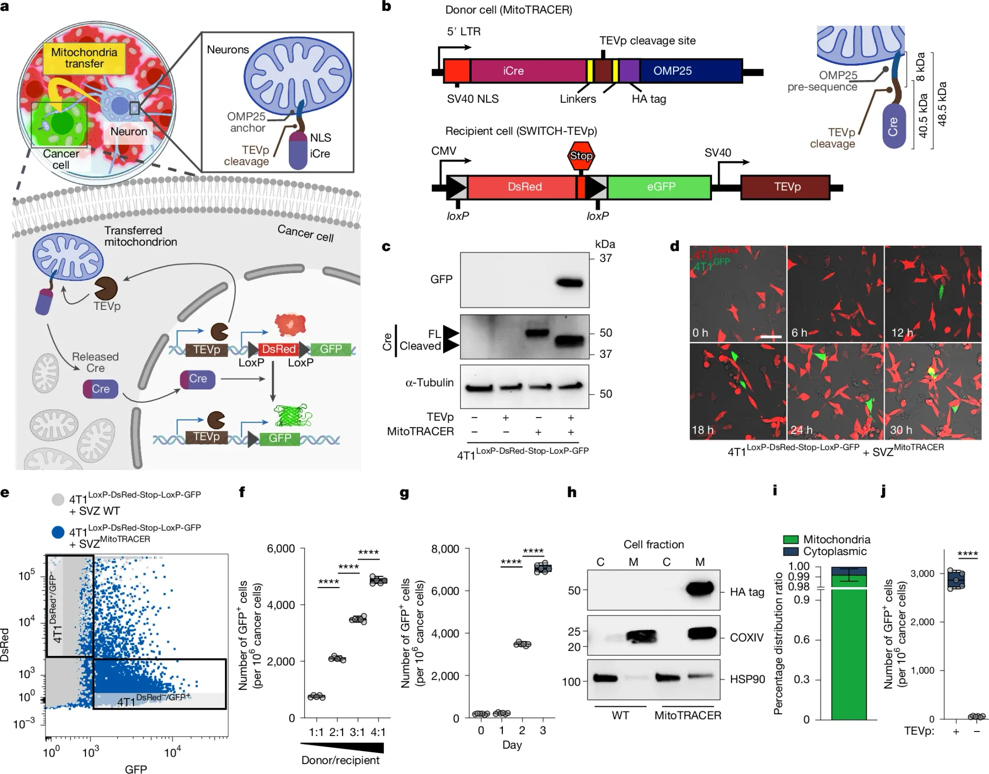
The researchers behind this week's paper built on this idea and developed a genetic tool called MitoTRACER to permanently label cancer cells that receive mitochondria from neurons.
This work can be seen in the figure above a) is a diagram of how the system works - mitochondria in donor neurons are labeled with Cre which is released by TEVp if those mitochondria are transferred into cancer cells that have a dsRed/GFP/TEVp expressing construct. If cancer cells don't get any neuronal mitochondria, they're red, if they do, Cre does its magic and cuts out the dsRed construct allowing the expression of GFP and turning the cells green! b) displays the constructs used in the donor neurons and the cancer cells c) shows that the system works using a western blot d) it works using confocal microscopy e) and with FACS f,g) shows that transfer is both concentration and time dependent and h,i) show that the MitoTRACER components are stuck to the mitochondria and not just floating around the cytoplasm.
Using this system in breast cancer models, it was shown that mitochondria are directly transferred from neurons to cancer cells.
And also that cancer cells that acquired these mitochondria gained enhanced OXPHOS capacity and resistance to metastatic stressors like oxidative damage and mechanical shear.
Further analysis of human tumors confirmed that cancer cells near nerves had greater mitochondrial content, and that denervated tumors had significantly lower mitochondrial load.
These findings solidify the idea that neuronal mitochondria serve as a critical metabolic resource for cancer cells during metastasis.
And if this is true, it means we can use this to our advantage therapeutically!
Because stopping mitochondrial transfer from neurons to tumor cells, inhibiting tumor-induced neurogenesis, or using denervation techniques may significantly impair the ability of tumors to spread and resist therapy.
###
Hoover G, et al. 2025. Nerve-to-cancer transfer of mitochondria during cancer metastasis. Nature. DOI: 10.1038/s41586-025-09176-8
Studying the microbiome is really just a lesson in ecology!
The word microbiome was first used to describe all of the microbes that inhabited a specific environment.
Many of the words we use when talking about the microbiome come from ecology which is the study of how organisms (usually plants and animals) interact with each other and their environments.
So, the microbiome is actually something that’s studied as a part of the field of microbial ecology which covers basically all of the different microbiomes.
We might like to think that “the microbiome” is a human centric thing, but that’s a surefire way to make a microbial ecologist angry!
Because plants, animals, the soil, and even your toilet seat have a microbiome!
In traditional ecology, a biome is a specific geographic area that is defined by the climate and the plants and animals that inhabit it.
But instead of talking about polar bears and walruses living in the arctic tundra, for the gut microbiome, we talk about the Prevotella, Bacteroides, and Clostridium that colonize your colon!
Today the microbiome is defined as the community of microorganisms that live on or in a specific environment and how they interact with one another.
These include bacteria, fungi and viruses but also the activities and interactions of their epigenomes, genomes, transcriptomes, proteomes and metabolomes.
Much like any other complex biological system, the only way to truly understand the microbiome is by looking at all of the major -omes!
This also helps us to understand what roles all of these organisms play within their community and how they survive in their niche.
Because just like in ecology, relationships exist between organisms and these can include:
Predation - Pretty straight forward, a predator eats prey.
Competition - A battle between two or more organisms over limited resources.
Commensalism - One organism benefits from a relationship and the other is unchanged.
Mutualism - Both organisms benefit from a relationship.
Parasitism - One organism benefits while the other is harmed.
So, why am I going into excruciating detail about ecology as it relates to the microbiome?
Because it's important to understand that if we want to learn anything about how we can alter the microbiome to improve human health we also need to realize that it's an incredibly complex biological system all in itself!
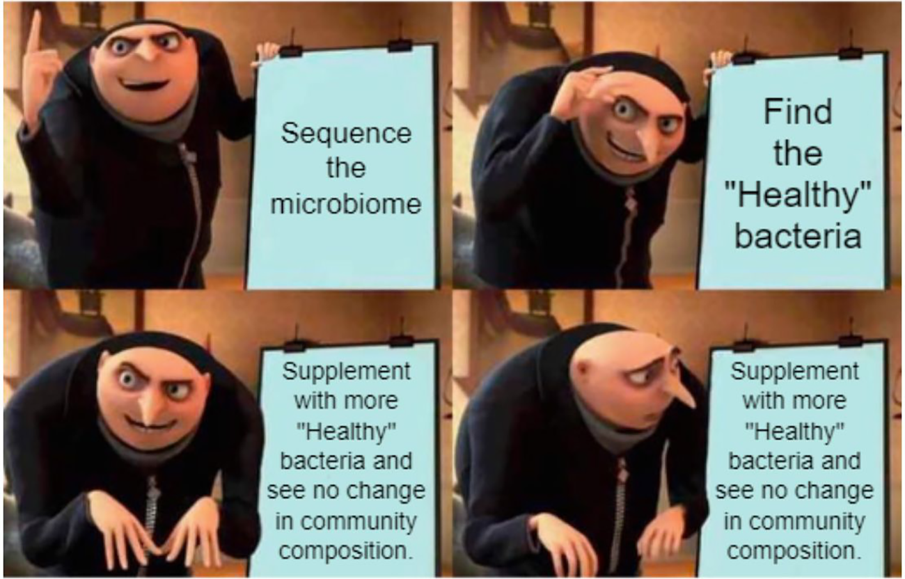
The microbiome isn't just about identifying the organisms in a community and saying “these are healthy and these other ones aren't.”
That's because the role of an organism changes in complex ways based on what other organisms are present and the environment in which it resides.
So, simple approaches and explanations of the microbiome won't do and there's still a lot of science that needs to be done before we can start changing the human microbiome to our benefit.
Watson and Crick solved the structure of DNA and everyone lived happily ever after, right? Wrong. That was just the opening argument.
Because once the structure was proposed, everyone needed something new to fight about.
And one of those new topics was how a double stranded DNA helix was copied or 'replicated' to then be passed on to the next generation.
There were three theories for how this might work:
Conservative - A completely new copy of DNA is made that contains no pieces from the parental molecules.
Dispersive - Pieces of the parental molecule are copied and interspersed with fragments of the daughter molecules so the replicated copy is a mishmash of old and new.
Semi-conservative - One strand of a parent molecule serves as a template for creating the daughter strand so one old strand and one new strand end up in the new helix.
Watson and Crick thought the semi-conservative model was the simplest and made the most sense.
But Max Delbrück thought that was impossible because it would require the helix to totally unwind.
He believed that random copying of parent fragments was more likely so he was the champion of the dispersive model.
While the heavyweights were arguing about their theories, Matthew Meselson and Franklin Stahl, two recently minted PhDs, decided to do some actual work.
So, in 1954, they designed a series of experiments to use density gradient ultracentrifugation to settle this latest quarrel.
Now, the method they used might sound complicated but really all they did was extract DNA and spin it really really hard for a very long time - like, 45,000 rpm hard and for 20 hours.
But centrifuging the DNA was only half of the solution, they needed to figure out a way to make ‘old’ DNA strands heavy and ‘new’ ones light.
After a bit of trial and error, they settled on growing bacteria in the presence of a heavy isotope of Nitrogen, N15 and then switching the growth media to regular N14 to see what happened.
If DNA replication was semi-conservative, and not dispersive (random), they hypothesized they would see very sharp bands after centrifugation as the lighter new strands mixed with the old heavy ones.
If there was no banding, and more of a smear, they’d know replication was dispersive.
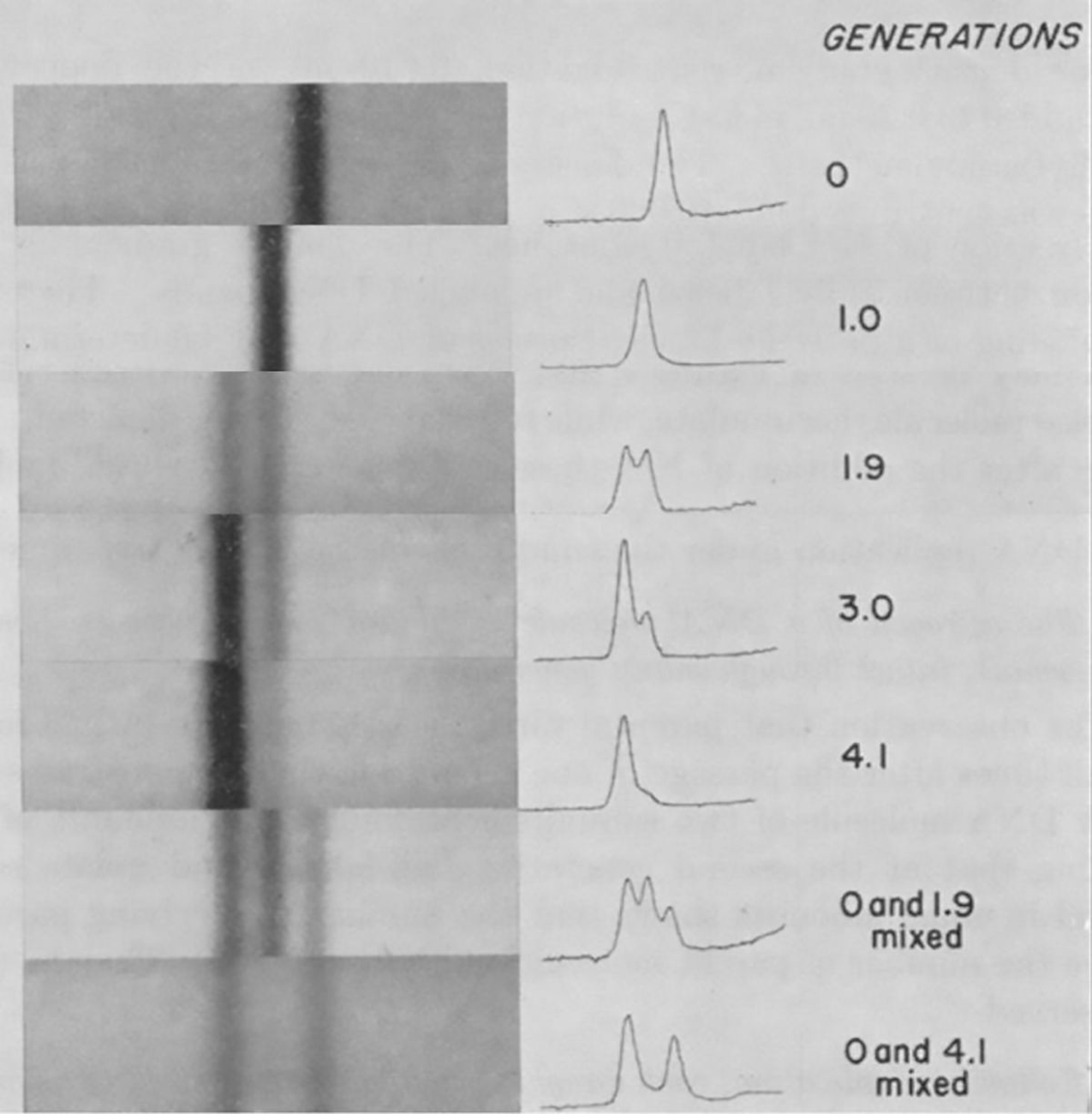
The results of this experiment are displayed in the figure above and are exactly what would be expected if DNA replicated semi-conservatively.
In Gen 0, everything is heavy, in Gen 1, the helix has one heavy and one light strand, in Gen 2, 50% of the helices are light and 50% are a mix, and by Gen 4, the majority of the DNA helices are composed of light strands.
This simple yet elegant experiment was all it took to convince Max Delbrück that he was wrong and it proved that DNA replication was semi-conservative.
###
Meselson M, Stahl FW. 1958. The replication of DNA in Escherichia coli. PNAS. DOI: 10.1073/pnas.44.7.671